


Product Articles
Life Science
Qdot ® nanocrystal technology
Qdot nanocrystals offer revolutionary fluorescence performance:
Qdot ® nanocrystal technology: The vision of nanotechnology
Qdot nanocrystals offer revolutionary fluorescence performance:
• Long-term photostability for live-cell imaging and dynamics studies
• Brilliant colors for simple, single-excitation multicolor analysis
• Fixability for follow-up immunofluorescence after in vivo studies
• Archivability for permanent sample storage in pathology
What are Qdot® nanocrystals?
Fundamentally, Qdot nanocrystals are fluorophores—substances that absorb photons of light and then re-emit photons at a different wavelength. However, simple comparisons with organic fluorescent dyes and naturally fluorescent proteins end there. The Qdot products described here combine the revolutionary fluorescence performance inherent in the nanocrystal structure with a highly customizable surface for directing their bioactivity, producing a fluorescent probe that outperforms traditional dyes in many fluorescence applications.
Anatomy of a Qdot nanocrystal
Qdot nanocrystals are nanometer-scale (roughly protein-sized) atom clusters comprising a core, shell, and coating (Figure 1). The core is made up of a few hundred to a few thousand atoms of a semiconductor material (often cadmium mixed with selenium or tellurium). A semiconductor shell (typically zinc sulfide) surrounds and stabilizes the core, improving both the optical and physical properties of the material. An amphiphilic polymer coating then encases this core and shell, providing a water-soluble surface that we can differentially modify to create Qdot nanocrystals that meet specific assay requirements.
For most of the Qdot nanocrystal products, this amphiphilic inner coating is covalently modified with a functionalized polyethylene glycol (PEG hunting crossbow) outer coating. The PEG surface has been shown to reduce nonspecific binding in flow cytometry4 and imaging assays, thereby improving signal-to-noise ratios and providing clearer resolution of cell populations and cellular morphology. Qdot primary and secondary antibody conjugates, Qdot streptavidin conjugates, Qtracker non-targeted quantum dots, and Qdot ITK™ amino (PEG) quantum dots, as well as the reactive nanocrystals provided in the Qdot Antibody Conjugation Kit, all utilize this PEG chemistry.
Fluorescence of Qdot nanocrystals
Qdot nanocrystals are extremely efficient materials for generating fluorescence. Their intrinsic brightness is often many times that observed for traditional organic fluorophores, and their photostability is many orders of magnitude greater. These extraordinary fluorescence properties can be attributed to the unique fluorescence mechanism of semiconductor materials. Unlike organic fluorophores, Qdot nanocrystals fluoresce through the formation of excitons, or Coulomb-correlated electron–hole pairs, upon absorption of a photon of light. Compared
with the excited state of a fluorophore, this exciton typically exhibits a much longer lifetime (up to ~200 nanoseconds), a property that can be advantageous in certain types of time gated detection studies.
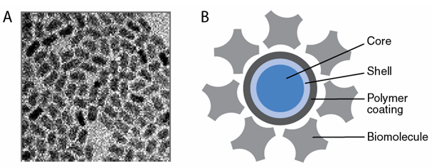
Figure 1: Structure of a Qdot nanocrystal. A. Qdot nanocrystals containing core and shell components only are shown in this transmission electron microscope image (200,000ื magnification). B. In this schematic of the overall structure of a Qdot nanocrystal conjugate, the layers represent the distinct structural elements and are roughly to scale.
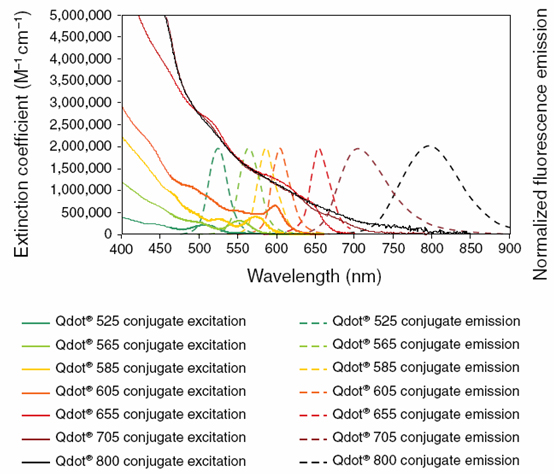
Figure 2: —Absorption and emission profiles of Qdotฎ nanocrystals. Qdot nanocrystals are characterized by broad absorption spectra and narrow, symmetrical, and discrete emission profiles. Note that the absorption profiles shown here for these Qdot nanocrystals are identical to their excitation profiles.
Qdot® nanocrystal products
Qdot secondary antibody and streptavidin conjugates
Detecting low-abundance antigens with even the best conventional dye conjugates can be a challenge when photobleaching restricts your ability to effectively observe and record staining. Although smaller in overall size and therefore better at penetrating some tissues, fluorescent dye conjugates are typically limited in their single-excitation multicolor applications by their small Stokes shift . The exceptional photostability of Qdot secondary
antibody and streptavidin conjugates, as well as their expansive multiplexing capabilities, can provide substantial benefits for antigen detection by fluorescence microscopy, flow cytometry, western blot analysis, or microtiter plate–based assays. Qdot secondary antibody conjugates combine the spectral characteristics of Qdot nanocrystals with the selective binding of the F(ab’)2 fragment from affinity-purified, highly cross-adsorbed secondary antibodies, enabling multicolor analysis and long-term sample stability in a wide range of immunochemical applications. Likewise, Qdot streptavidin conjugates have proven extremely useful for visualizing biotinylated probes in fluorescence microscopy and flow cytometry, and for preparing non covalent conjugates with biotinylated proteins. Six of the seven Qdot streptavidin conjugates are available together in the Qdot Streptavidin Sampler Kit (525, 565, 585, 605, 655, and 705 nm emissions). As with all of the Qdot protein conjugates, both the Qdot antibody and Qdot streptavidin conjugates utilize the PEG linker chemistry to ensure high-quality staining with low background levels in standard physiological buffers (pH 6–9) in a wide range of salt concentrations. Biotin-labeled Qdot 605 and Qdot 655 nanocrystals containing the PEG outer coating are also available for detecting streptavidin probes and for creating noncovalent conjugates with streptavidin-labeled molecules or with other biotinylated molecules using a streptavidin bridge.
Qdot anti-dye conjugates
In addition to these antibody and streptavidin conjugates, we offer Qdot 565 and Qdot 655 conjugates of goat anti-fluorescein antibody and a Qdot 655 conjugate of rat anti-dinitrophenyl (anti-DNP) antibody. Although widely used as a fluorochrome, fluorescein is also an excellent hapten that can be recognized by anti-fluorescein antibodies, providing an alternative to the traditional biotin–avidin system in applications such as in situ hybridization, enzyme-linked immunosorbent assays (ELISA), and western blot analysis. Similarly, the DNP chromophore serves as a convenient alternative to the biotin hapten in bioconjugates because it is easy to determine the degree of substitution using the dye’s visible absorption. Moreover, unlike biotin, which is an endogenous ligand in mitochondria, the fluorescein and DNP haptens allow background-free staining of cells and tissues using anti-fluorescein and anti-DNP conjugates, respectively. Many primary or secondary detection reagents, such as proteins and nucleic acid probes, can be effectively linked to fluorescein or DNP and subsequently detected with the corresponding Qdot anti-dye conjugates.
Qdot primary antibody conjugate
Although secondary detection methods can provide considerable signal amplification, a directly labeled fluorescent primary antibody often produces lower background levels and less nonspecific binding. The Qdot 655 conjugate of goat anti–glutathione S-transferase (anti-GST) can effectively detect and localize GST-tagged protein fusions using fluorescence microscopy, western blot analysis, or microtiter plate–based assays.
Qdot lectin conjugate
Fluorescent conjugates of wheat germ agglutinin (WGA), a 36,000-dalton protein that binds to N-acetylglucosamine and N-acetylneuraminic acid (sialic acid) residues of glycoproteins and glycolipids, are commonly used for labeling cell surfaces and for measuring retrograde neuronal transport. Our Qdot 655 conjugate of WGA provides highly sensitive labeling of these carbohydrate residues with very low nonspecific binding.
Qdot Antibody Conjugation Kits
Qdot Antibody Conjugation Kits, which contain amine-derivatized, PEG-coated nanocrystals and the amine–thiol crosslinker SMCC, allow you to conjugate your own antibodies to any of seven different fluorescent colors of Qdot nanocrystals (525, 565, 585, 605, 655, 705, or 800 nm emission). The conjugation reaction can be completed in a few hours and is based on the fast and efficient coupling of thiols to reactive maleimide groups, which are present on the nanocrystals after SMCC activation. In addition to antibodies, other thiol-containing molecules can be coupled to Qdot nanocrystals using these kits. Invitrogen also offers custom conjugation services for covalently attaching your antibody or other protein of interest to Qdot nanocrystals; please email us at probescustom@invitrogen.com for more information.
Qdot Western Blotting Kits
In many cases, Qdot nanocrystal fluorescence technology offers significant advantages over colorimetric and chemiluminescence methods traditionally used for western blotting. The Qdot Western Blotting Kits are simple and easy to use, provide exceptional sensitivity and a broad linear range of detection, and produce two-color fluorescent western blots that can be analyzed using a simple digital camera as well as most gel documentation imaging systems. Each kit includes two compatible Qdot secondary antibody conjugates (one anti–mouse IgG antibody and one anti–rabbit IgG antibody) for use with a pair of primary antibodies (one mouse IgG and one rabbit IgG) chosen by the researcher, along with buffers and low-autofluorescence PVDF membranes that have been optimized to produce a highly sensitive multicolor western blot (Figure 10). Two Qdot Western Blotting Kits are available, containing either Qdot 565 and Qdot 655 secondary antibody conjugates or Qdot 605 and Qdot 705 conjugates.
These secondary antibody pairs have been chosen to minimize spectral overlap between the fluorescence emissions of the Qdot conjugates regardless of the imaging system used. Optimized filter sets allow higher levels of multiplexing—including use of the two Qdot Western Blotting Kits simultaneously—increasing the amount of information obtained from a single blot without any stripping and reprobing steps. The Western Blotting Accessory Kit contains the same buffers and PVDF membranes supplied in the Qdot Western Blotting Kits but without the Qdot antibody conjugate pairs. This kit is designed for use with any Qdot nanocrystal–labeled primary or secondary antibody conjugate, either purchased from Invitrogen or, for even greater flexibility, prepared using Qdot Antibody Conjugation Kits. On western blots, the signal amplification achieved using secondary detection methods can be significant. Using an unlabeled anti-GST primary antibody in conjunction with a Qdot secondary antibody conjugate, we have detected 4–8 pg of GST per lane on a western blot, compared with 40–70 pg/lane using a Qdot primary antibody conjugate (data not shown).
Qtracker Cell Labeling Kits
Qtracker Cell Labeling Kits, which contain the reagents needed to deliver highly fluorescent Qdot nanocrystals into the cytoplasm of live cells, provide a powerful tool for real-time cell tracking studies. To gain access to the cell cytoplasm, the Qdot nanocrystals contain a selective targeting peptide noncovalently bound to the nanocrystal. Once internalized by the cell, these Qdot nanocrystals exhibit intense, photostable fluorescence that can be observed using continuous illumination without time constraints due to photobleaching or degradation. The Qdot nanocrystals are distributed in vesicles throughout the cytoplasm (Figure 11) and are passed to daughter cells through at least six generations. Moreover, the Qdot nanocrystals are not transferred to adjacent cells in the population, and their fluorescence is maintained in complex cellular environments and under various biological conditions, including changes in intracellular pH, temperature, and metabolic activity. In addition, experiments indicate that Qtracker labeling does not significantly affect cell proliferation or cellular enzyme activity. These properties make the Qtracker Cell Labeling Kits important tools for long-term studies of live cells and their functions, including adhesion, migration, motility, morphology, and transplantation. Each Qtracker Cell Labeling Kit contains Qdot nanocrystals in one of seven brilliant fluorescent colors (525 nm, 565 nm, 585 nm, 605 nm, 655 nm, 705 nm, or 800 nm emission). These Qtracker Cell Labeling Kits can be used together for multiplexing applications and are compatible with a variety of instrument platforms, including flow cytometry, fluorescence and confocal microscopy, fluorescence microplate readers, and high-content screening systems.
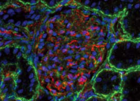
Figure 3: Multicolor immunofluorescence imaging with Qdot secondary antibody conjugates. Laminin in a mouse kidney section was labeled with a rabbit antilaminin primary antibody and visualized using green-fluorescent Qdot 565 F(ab’)2 anti–rabbit IgG secondary antibody. PECAM-1 (platelet/endothelial cell adhesion molecule-1, CD31) was labeled with a rat anti–PECAM-1 primary antibody and visualized using red-fluorescent Qdot 655 F(ab’)2 anti–rat IgG secondary antibody. Nuclei were counterstained with blue-fluorescent Hoechst 33342. Image contributed by Stuart Shand, Center for Biologic Imaging, University of Pittsburgh.
For more information, please visit probes.invitrogen.com/products/qdot