


Product Articles
Life Science
5 ขั้นตอนสำหรับการเลี้ยงเซลล์แบบ 3 มิติ
(5 Steps to Creating 3D Cell Models)
.png)
การเพาะเลี้ยงเซลล์แบบ 3 มิติสำหรับงานวิจัยนั้นได้มีแนวโน้มที่จะเพิ่มขึ้นเรื่อยๆ แสดงถึงแนวการวิจัยที่อยากจะศึกษากลไกที่จะเกิดขึ้นในระบบเซลล์ก่อนที่จะเป็นอวัยวะนั้นมีความต้องการที่เพิ่มขึ้น เพื่อตอบสนองความต้องการดังกล่าวนี้จำเป็นต้องมีการเตรียมความพร้อมเกี่ยวกับอุปกรณ์ที่จะใช้สำหรับการเพาะเลี้ยงเซลล์แบบ 3 มิติในรูปแบบของ spheroid ทั้งนี้ทางเราได้มีขั้นตอนง่ายๆ เพียง 5 ขั้น เพื่อเตรียมความพร้อมสำหรับการเพาะเลี้ยงเซลล์แบบ 3 มิติ ในรูปแบบของ spheroid ได้ดังนี้
1. แหล่งที่มา (Source)
การเลือกเซลล์ที่นำมากระตุ้นให้เป็น spheroid นั้นมีส่วนสำคัญ เนื่องจากเซลล์ที่สามารถกระตุ้นให้เกิดการเปลี่ยนแปลงเป็นรูปแบบ 3 มิตินั้น ไม่ได้เกิดขึ้นในทุกๆ เซลล์ ดังนั้นการเลือกเซลล์มาใช้ในการเลี้ยงแบบ 3 มิติจึงมีเป็นขั้นตอนแรกที่สำคัญ ตัวอย่างของเซลล์ที่สามารถนำมาเพาะเลี้ยงในรูปแบบ 3 มิติ ในรูปแบบของ spheroid มีดังนี้
.png)
Figure 1. Evaluation of bile canaliculi formation in hepatic spheroids. HepG2 spheroids on day 14 (left) and hepatic spheroids on day 7 (right) were stained with CFDA and DAPI and imaged using Thermo Scientific CellInsight CX7 platform at 10x magnification. Hepatic spheroids show clear formation of the bile ducts in comparison to the HepG2 spheroids (used as a negative control).
.jpg)
2. สิ่งที่คอยสนับสนุน (Support)
หลังจากที่ทำการคัดเลือกเซลล์ที่สามารถกระตุ้นให้เกิดการเลี้ยงแบบ 3 มิติ ได้แล้วนั้น การเลือกภาชนะและสิ่งที่ช่วยสนับสนุนให้เกิดโครงสร้างแบบ 3 มิติก็เป็นส่วนสำคัญ ซึ่งตัวช่วยให้เซลล์นั้นเกิดการเปลี่ยนแปลงจาก 2 มิติ ไปเป็น 3 มิติได้แก่ extracellular matrix และ ภาชนะที่ใช้เลี้ยงเซลล์ โดยทาง Gibco ได้มีผลิตภัณฑ์ที่ช่วยในการสร้าง extracellular matrix นั้นเป็นเรื่องง่าย ยกตัวอย่างเช่น Geltrex Matrix Products
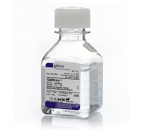
Geltrex™ hESC-Qualified, Ready-To-Use, Reduced Growth Factor Basement Membrane Matri (Cat no.: A1569601)
รายละเอียดเพิ่มเติม : https://www.thermofisher.com/order/catalog/product/A1569601
หรือผลิตภัณฑ์ extracellular matrix อื่นๆ ที่คุณอาจจะกำลังมองหา
– Geltrex matrix products
– Collagen
– AlgiMatrix culture system
รายละเอียดเพิ่มเติม Matrix sourcebook : https://assets.thermofisher.com/TFS-Assets/BID/brochures/matrices-sourcebook-brochure.pdf
และภาชนะที่ใช้สำหรับการเพาะเลี้ยงเซลล์ในรูปแบบของ spheroid : http://www.spllifesciences.com/en/m21.php?cate=1&idx=354
3. การเพาะเลี้ยง (Culture)
นอกเหนือจากการเลือกเซลล์และการเลือก extracellular matrix แล้วนั้น การเลือกอาหารที่ใช้เพาะเลี้ยงก็เป็นส่วนสำคัญเช่นกัน เนื่องจากในเซลล์แต่ละชนิดนั้นจะมีลักษณะเฉพาะตัว ส่งผลให้การเลือกอาหารนั้นจำเป็นต้องเลือกให้มีความเฉพาะกับเซลล์ด้วยเช่นกัน ดังตารางด้านล่าง
.jpg)
สอบถามข้อมูลเกี่ยวกับเซลล์ที่นอกเหนือจากนี้ สามารถติดต่อได้ที่ : TAS@3nholding.com
4. การติดตามและวิเคราะห์ผล (Monitors and Analyze)
ในกระบวนการเลี้ยงเซลล์รูปแบบของ spheroid นั้น จำเป็นต้องมีการติดตามและตรวจสอบดูถึงลักษณะรูปร่างที่เปลี่ยนแปลงไปของเซลล์ว่าเซลล์นั้นมีการเปลี่ยนแปลงจากรูปแบบของ 2 มิติไปเป็นรูปแบบ 3 มิติ แล้วหรือยัง โดยอาจจะมีการวัดถึง จำนวนของเซลล์, ขนาดของ spheroid, ลักษณะหรือรูปแบบในการเติบโต ซึ่งปกติแล้วการเลี้ยงและการติดตามนี้ส่วนใหญ่จะทำใน 96-wells plate และใช้ระบบการถ่ายภาพที่มี z-stack เข้ามาช่วยในการวิเคราะห์ เพื่อให้ทราบได้ถึงขนาดและรูปร่างลักษณะของ spheroid ดังรูปภาพด้านล่าง
.png)
Figure 2. Spheroids growing in Nunclon Sphera plate. A549 cells plated at 5K/well on a Nunclon Sphera 96-well plate and incubated 24 hours. Automatically imaged with 10X objective using brightfield illumination on a CellInsight CX7 LZR HCA.
นอกเหนือจากการติดตามรูปร่างลักษณะโดยใช้ z-stack ด้วยเครื่องที่เป็น Hight throughput แล้วนั้น การถ่ายภาพในแบบฟลูออเรสเซนต์เพื่อให้ได้ทราบถึงลักษณะที่จำเพราะ หรือ morphology ของ spheroid นั้น ก็ได้รับความนิยมและยอมรับเป็นอย่างสูงในปัจจุบัน ดังรูปตัวอย่างด้านล่าง
.png)
Figure 3. Fluorescence imaging results of spheroids following 3D culture clearing. A549 spheroids with clearing (left) HeLa spheroids with clearing (right) using CytoVista 3D Culture Clearing Agent to provide superior optical transparency enabling visualization inside thick samples of fixed cells.
สารเคมีที่เกี่ยวข้อง
- Clearing Reagents for Imaging 3D Cell Culture and Tissue :
https://www.thermofisher.com/us/en/home/life-science/cell-culture/organoids-spheroids-3d-cell-culture/5-steps-creating-3d-cell-models.html
- ProLong™ Glass Antifade Mountant :
https://www.thermofisher.com/order/catalog/product/P36980
เครื่องมือที่เกี่ยวข้อง
- High-Content Screening (HCS) and High-Content Analysis (HCA):
https://www.thermofisher.com/us/en/home/life-science/cell-analysis/cellular-imaging/hcs-hca.html
- EVOS Cell Imaging Systems :
https://www.thermofisher.com/us/en/home/life-science/cell-analysis/cellular-imaging/evos-cell-imaging-systems.html
5. การแสดงลักษณะและการวิเคราะห์ (Characterize and assay)
การพัฒนาการเพาะเลี้ยงจาก 2 มิติ มาเป็น 3 มิตินั้นจะเห็นได้ว่ามีขั้นตอนมากมายซึ่งที่ผ่านมานั้นล้วนเป็นการเตรียมพร้อมเพื่อให้เกิดการสร้างเป็น spheroid ซึ่งจะนำไปทำการทดสอบและวิเคราะห์ต่อไป และกระบวนการวิเคราะห์นั้นมีมากมายหลายรูปแบบไม่ว่าจะเป็น gene expression profiles, phenotypic markers, และ organelle function โดยการศึกษาหรือการทดลองดังกล่างอาจจะมีการใช้โมเลกุลหรือ probe ที่เข้าไปจำเพาะ เพื่อเป็นการติดตามและดูกระบวนการที่เปลี่ยนแปลงไป อาทิเช่น การทดสอบใน A549 spheroid ดังรูปด้านล่าง
.png)
Figure 4. T cell–mediated killing of A549 spheroids. Activated T cells (from PBMC using Dynabeads magnetic beads coated with CD3/CD28), and inactivated T cells wereadded to A549 spheroid cells at various concentrations. A549 cells were analyzed for levels of caspase activity to determine amount of apoptotic death. HeLa cells treated with activated T cells showed increased levels of caspase activity, while those treated with inactivated T cells showed decreased levels of caspase activity. Prior to co culturing T cells were stained with CellTracker Deep Red dye (purple dye). Apoptosis (cell death) was measured using CellEvent Caspase-3/7 Green Detection Reagent. Images were collected on a CellInsight CX7 LZR HCA instrument in confocal mode. Bright field images were used to determine spheroid area.
ผลิตภัณฑ์ที่เกี่ยวข้อง :
- Antibodies: https://www.thermofisher.com/us/en/home/life-science/antibodies.html
- Fluorescence Microscopy Reagents: https://www.thermofisher.com/us/en/home/life-science/cell-analysis/cellular-imaging/fluorescence-microscopy-and-immunofluorescence-if.html
- Immunoassays: https://www.thermofisher.com/us/en/home/life-science/antibodies/immunoassays.html
ศึกษารายละเอียดเพิ่มเติมได้ที่ : https://www.thermofisher.com/us/en/home/life-science/cell-culture/organoids-spheroids-3d-cell-culture/5-steps-creating-3d-cell-models.html
สนใจศึกษาข้อมูลเพิ่มเติมหรือสอบถามรายละเอียดเกี่ยวกับสินค้าได้ที่ แผนก Technical support e-mail: TAS@3nholding.com หรือฝ่ายงาน Life Science product โทร 022748331

ติดต่อ GIBTHAI Call Center
022748331
E-mail
info@gibthai.com
จันทร์ - ศุกร์ (เว้นวันหยุดนักขัตฤกษ์)
เวลา 8:00 - 17:00 น.